|
Are we to blame?
Could it really be the case that with the recent research
that has been done into tooth problems in domesticated Chinchillas, that the
feed we give our animals and the stress that they have in their lives as
pets is in part the reason as to why so many pet Chinchillas suffer from tooth
disease?
Read the evidence and findings for yourselves,
of the studies that were undertaken in the last few years to find out more.
As always the Chinchilla Club would love to hear your views
on this subject and the research findings, please
do get in touch!
Forward written by - Anjela Ross
For - The Chinchilla Club
|
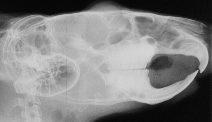
This chinchilla is 'clinically' normal .
|
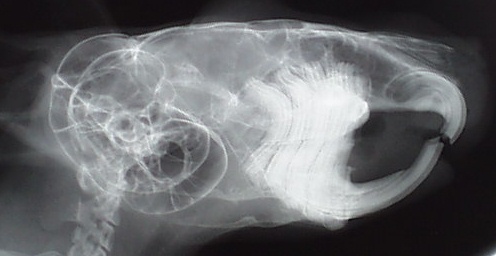
This x-ray shows crown and root elongation with obvious
root deformity in a chinchilla.
|
|
THE SITE OF OBSTRUCTION OF THE LACRIMAL DRAINAGE
SYSTEM IN CHINCHILLAS (Chinchilla lanigera) WITH "wet eyes"
Research by - Crossley DA, Roxburgh G
(1999)
Introduction:
A common clinical problem seen in chinchillas (Chinchilla lanigera)
is "wet eyes" due to lacrimal overflow.
Aim:
To investigate the site or sites of obstruction of the lacrimal
drainage system in chinchillas showing signs of lacrimal overflow.
Method:
Radiography, CT scanning, anatomical dissection, and histological
examination of affected animals. signs of lacrimal overflow.
Results:
The main site of obstruction of lacrimal drainage is in the
descending portion of the lacrimal canal between the orbit and the incisor
tooth root apex. Bony remodelling around elongating maxillary premolar and
first two molar tooth roots intrudes into the lacrimal canal compressing
and sometimes occluding or even obliterating the lacrimal canal and duct.
No evidence has been found for obstruction adjacent to the incisor root
apices in the specimens examined so far.
Discussion:
Lacrimal drainage in healthy chinchillas is similar to that
in other rodents. This species is adapted to a highly abrasive herbivorous
diet, having continuously growing cheek teeth (in addition to the continuously
growing incisors). When these teeth are not worn adequately, i.e. when domestic
animals are fed commercial diets, the teeth continue to elongate. Eventually
occlusal pressure prevents eruption, so the roots intrude inducing remodelling
of adjacent tissue including the lacrimal canal.
Conclusions:
Chinchillas should be fed a herbivorous diet which requires prolonged
chewing in order to wear the teeth adequately.
|
|
DENTAL DISEASE IN CHINCHILLAS IN THE UK
David A. Crossley
Dental abnormalities are common in chinchillas, however,
knowledge of the nature and relative incidence of the lesions responsible
for clinical signs is incomplete. Animals, radiographs and/or prepared specimens
were examined to gain further knowledge regarding dental anatomy and dental
disease in the UK chinchilla population.
Thirty five percent of the apparently healthy chinchillas
examined had dental abnormalities detectable on routine examination. The
range and relative incidence of different dental abnormalities encountered
in these and clinically affected animals are presented. Whilst malocclusion
was a common finding, in all but one case this was secondary to crown elongation
or absence of opposing teeth, not a primary skeletal problem.
Clinical signs commonly attributed to malocclusion, such
as ventral mandibular swelling, weight loss, dysphagia, altered chewing
pattern and changed food preferences, are not specific to malocclusion.
They are seen associated with tooth root elongation, spike formation on
the sides of the occlusal surfaces and advanced periodontal lesions. Caries
and odontoclastic resorptive lesions rarely cause obvious clinical signs
but were identified regularly. Congenital absence of cheek teeth, true skeletal
malocclusion and pathological loss of incisor teeth all resulted in significant
clinical signs but were rarely seen.
Lack of dietary abrasion and stress are probably the main
aetiological factors for the most prevalent dental problem, cheek tooth
elongation. Combination of provision of a diet matching that eaten by wild
animals and reducing stress levels of captive chinchillas should reduce
the incidence of dental disease in this species.
|
|
SKULL SIZE AND CHEEK TOOTH LENGTHS IN WILD
AND CAPTIVE CHINCHILLA POPULATIONS
David A. Crossley and Maria del Mar Miguélez
(2001)
Summary:
Chinchillas are herbivorous rodents with teeth that all grow
continuously. In captivity they are commonly affected by dental disease.
Since the range of dental disease occurring in wild chinchillas is unknown,
the dentition of museum specimens originally obtained from the wild was
assessed and compared with specimens prepared from captive bred animals.
Skulls from wild-caught chinchillas showed minimal evidence
of dental disease and the teeth were all short, cheek tooth lengths averaging
5.9 mm. Cheek tooth lengths in zoo specimens (average 6.6 mm), clinically
normal (average 7.4 mm) and captive bred animals with dental disease (average
10 mm) were significantly elongated by comparison (p < 0.0001). Captive
bred specimens showed a wide range of tooth related lesions.
These results suggest that some aspect of captivity is
responsible for the development of dental disease in chinchillas. It is
suggested that the diet (its physical form and composition) is the main
etiological factor, and that provision of a diet closely matching that of
wild chinchillas should significantly reduce the incidence of dental disease
in captive chinchillas.
|
|
|
